Anatomy & Disorders OF Knee

Anatomy And Disorders Of The Knee The human body consists of many joints that also vary in type. A joint is defined as the articulation of any two or more bones. Different types of joints are so classified depending upon the range of motion they permit. For example, Shoulder joint allows complete rotation of the arm whereas the Elbow joint allows only flexion and extension. The above-mentioned example exhibits how two different joints on the same part of body (arm) permit different range of motion. In case of the shoulder, rotation of the arm is possible because the head of humerus (bone in the upper part of arm) articulates with the socket of shoulder girdle. The socket formed by Glenoid Cavity and the articulating ball in the form of head of humerus make up the ball and socket joint that permits motion along the entire circumference because of the ball moving in all directions inside the socket. Similarly, the elbow joint functions like a hinge used in door. Just the way a hinge permits motion of the door/window along only one line, similarly the elbow joint permits only flexion and extension (along one line) motions. There are other types of joints too but it is the hinge joint that we would concentrate on for a simple reason that our joint-of-interest is Knee and it happens to be of hinge type.
A Normal Knee The above discussion tends to undermine the complexity of a joint as it talks only about two bones moving in relation to each other and thus producing the motion. The actual anatomy is much more complex as it involves all the soft tissue in and around the joint that may/may not aid in bringing about the motion. Besides the bones, a joint also consists of muscles, tendons, ligaments, cartilage etc. Besides the tissues mentioned, there are other connective tissues like Synovium that also form a part of the joint. Above all these, there always is the vascular and nervous supply that spans the entire human body and is as important as any other structure/organ/tissue inside the body. For a joint to function all these tissues are required because a bone cannot move on its own. The movement is brought about by contraction and relaxation of muscle fibres. To enable the muscles to contract or relax, vascular and nervous supply is a must. To facilitate the movement and to maintain the range of motion without bringing about the wear and tear of the articulating bones, we need the soft tissues like Cartilage and Synovium. Ligaments and Tendons bring about the firm attachment of either bone-to-bone or muscle-to-bone and provide stability to the joint over entire range of motion. Having appreciated the importance of other tissues present inside the joint, we will proceed to learn which tissues are present inside the Knee joint and what role each of them performs. Though this unit will try to cover all the tissue of importance in Arthroscopy, it will not be exhaustive and further reading of the subject from other literature available is strongly recommended.
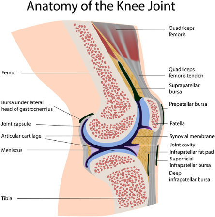
Bones of the Knee Joint The Knee joint is made up of 4 bones; namely Femur, Tibia, Fibula and Patella. Femur, the longest bone of the body, articulates in the knee joint at its distal end and it is the proximal end of Tibia and Fibula that articulates with the Femur in Knee. Patella is the Knee cap. It should be noted that though the distal end of Femur and proximal end of Tibia articulate, they are not exactly complimentary to each other. The Femur ends distally into two protruding globes known as Condyles. Condyles are cancellous in nature and covered with Hyaline cartilage at their tips to minimise the friction when they glide on the Tibial surface. The femoral condyles form a deep groove called Intercondylar Notch between themselves. This notch is a very important landmark in the joint because through it pass the ACL and PCL. As mentioned, Tibia has a plateau-like surface at its proximal end that articulates in the knee joint. This Tibial plateau accommodates the Condyles of Femur. To accommodate Femoral Condyles, Tibial plateau has two concave Condyles. These condyles cover most part of the Tibial plateau. Of the two, medial Tibial Condyle is more concave, longer in anterior-posterior direction and more oval while lateral Tibial Condyle is flatter and more rounded. This is to accommodate the bigger medial Femoral Condyle and rounder lateral Femoral Condyle. The other bone of the calf, i.e. Fibula is comparatively much weaker and runs lateral to Tibia. Patella is a seed-shaped bone suspended in the Femoral Quadriceps Tendon. On its posterior surface, Patella has 7 grooves that come in contact with Femur during flexion and/or extension.
Ligaments of the Knee Joint
The movement that occurs between Femur and Tibia is called as Translation in which the Femur glides over the Tibia. During translation, the Knee joint is stabilised by primary as well as secondary restraints. The primary support is provided by various ligaments that attach themselves to one or the other bone of the joint whereas the secondary support comes from capsule & muscles around the joint. Primary support is provided by 5 ligaments as mentioned below:
Anterior Cruciate Ligament
Posterior Cruciate Ligament
Lateral Collateral Ligament
Medial Collateral Ligament
Coronary Ligament
The location and function of each of these ligaments is given in the following paragraph. The lateral and the medial collateral ligaments are present on either side of the joint to restrict medial-lateral movement between femur and tibia. Lateral collateral ligament (LCL) extends from the lateral side of femur to the lateral side of head of Fibula. Medial collateral ligament (MCL) extends from medial femur to the medial aspect of tibia. Thus, among themselves, these two ligaments take care of medial-lateral stability of the knee joint.
Menisci in the Knee Joint
Apart from the ligaments mentioned above, the knee is also stabilised by special structures of type fibrocartilage, known as Menisci. Menisci are two in number and present on the tibial plateau. These menisci have a peculiarity that they are tough and compact like cartilage where they are compressed between the bones but fibrous and flexible at their attachments. They rest between the femoral condyles and the tibial plateau in order to absorb shock during the use of joint, help to protect the articular cartilage-covered condyles and at the same time stabilise the joint. Each meniscus is named depending upon the side it is. Thus we have a medial meniscus and a lateral meniscus in each knee. The menisci attach themselves to the plateau at their ends, commonly referred to as ‘horns’. In addition to these attachments, they are also attached to the underlying cartilage along its circumference by Coronary Ligament.
Other Soft Tissue in and around the Knee Joint
Besides the ligaments, the knee is also stabilised by the capsule and muscles in around the joint. Out of these, it is more important for us, as sales personnel, to know what Capsule is and what is it formed of. Muscles are also important but more so for the surgeons who operate on the joint. On learning the anatomy of knee joint, it is clear that there is a lot of empty space in the joint. This space is called as the Joint Space. It should be understood that this space does not remain uniform throughout the range of motion. It is essential for the space not to remain uniform as this provides the flexibility required throughout the range of motion. The shape of this space keeps on changing with varying degrees of flexion or extension. Also, the boundaries of the joint space are not continuous, if only the above mentioned soft tissues are considered. The boundaries are made up of the articulating bones, ligaments, menisci and even a few muscles. Still, there are quite a few places where there is no soft tissue. To make the boundaries entire, a special connective tissue called Synovium is present inside the knee joint. Thus, Synovium aids in completion of the entirety of the Capsule. Besides this, the Synovium also performs the following three functions: a) fluid and colloid transport in and out of the joint, b) lubricate the joint and c) joint debridement by phagocytosis. The joint space is normally filled with Synovial fluid that is a form of plasma and contains the same electrolytes and antibodies found in plasma. Thus, in addition to lubricating the joint, Synovial fluid also nourishes the articular cartilage.
Disorders of the Knee
Disorders associated with the knee are numerous and not all of them are treated using minimally invasive technique. Of those treated using Arthroscopy, disorders can be classified as chronic or acute. It should be noted that all the disorders of the body can be broadly classified under these two heads for a simple reason that this classification is done on the basis of time. Chronic implies continued over long periods of time and acute means in one instant. Thus, fracture caused due to accident will be classified as acute injury whereas weakness of bone due to osteoporosis will be classified as chronic defect. Osteoporosis cannot occur in a flash. It is a result of bone quality degradation over a period of time. Similarly, in the Knee joint, there are some disorders that happen in a flash whereas others.
Of the acute injuries in the knee, most common is the meniscal tear followed by ACL rupture. Note that though these are very common, they are common only in highly active people who use the joint to the fullest. Frequent sufferers are sportsmen/women who play Football, Basketball, Hockey and other similar physically demanding games. As mentioned earlier on, 80% of the injuries of menisci occur in the medial meniscus. A meniscus gets injured because it gets impinged in between the articulating bones. Naturally, the type of injury is ‘tear’. As a thumb-rule, tears in the white-white zone are treated by resecting the meniscus, in the red-red zone are treated by repairing/stitching the meniscus while those in the red-white zone are upto the discretion of the surgeon depending upon whether the tear will heal or not. This thumb rule is normally followed because tears in red-red zone can heal on repair because of the vascularisation. As white-white is completely devoid of vascular supply, it will not heal even if repaired.
These tears are also classified depending upon their orientation and severity. The details of different types of tears can be read from the module supplied by Endosys. It should be noted that it is very important to know different types of tears because only then can one know how to treat it. Even in case of resection, when using our wide range of hand held instruments, there are different instruments for different parts of the meniscus and different types of tears. Application of different types of instruments for different zones of meniscus and different tears will be dealt with in a unit dedicated to Geofix hand held instruments.
Besides meniscal tears and ACL rupture, the other disorders that occur in the Knee are PCL, MCL and LCL rupture & Synovitis. Generally, PCL rupture is accompanied by rupture of ACL also. PCL in itself is a very strong ligament and is also placed such that it rarely faces strain enough to damage it. If such a strain does occur, it usually does simultaneously/after the rupture of ACL. Synovitis is nothing but inflammation of Synovium. This occurs because of infection and manifests itself as severe pain in the knee. The only remedy for this problem is resection of Synovium which is affected.
Equipped with the knowledge of anatomy and disorders of the Knee, we can now move on to the equipment used for Diagnostic Arthroscopy and Operative Arthroscopy.
Copyright © ReifierSoft 2017. All rights reserved